Keynote Forum
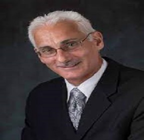
Title: Preventing Complications in Daily Dental Practice with Digital Measured Occlusion Technology
Abstract:
Former Assistant Clinical Professor, Department of Restorative Dentistry, Tufts University School of Dentistry, Boston, MA 02111, USA
Occlusion is a complex neurologic science that involves the interplay between teeth, muscles and the Temporomandibular Joints. Occlusion however, is taught biomechanically, absent of true interocclusal functional measurements of the occlusal force and timing of the contacts that affects patient’s neurologic health and comfort every time they use their teeth/prosthesis. The T-Scan 10 technology makes it possible to precisely control the occlusal forces and timing with natural teeth, dental implants, and prosthodontic restorations, offering patients predictably improved outcomes, while minimizing frequent common post-insertion occlusal adjustments that often don’t resolve the patient’s comfort issues, despite the dentist’s best efforts.
This course will present Digital Measured Occlusion treats common occlusal complications that appear in everyday clinical practice. Digital occlusion metrics guide the clinician to deliver comfortable, successful restorations, while eliminating the hit or miss subjectivity of using articulating paper, and overcoming the occlusal adjustment limitations that intraoral scanners and virtual articulation have during case insertion. And, because T-Scan accurately treats patients’ occlusal problems in MIP1-15, splints are less necessary. Clinical cases will illustrate how digitally diagnosing and treating occlusal problems to high-precision tolerances improves occlusal outcomes.
Figure
Biography:
Dr. Robert B. Kerstein received his D.M.D. degree in 1983, and his Prosthodontic certificate in 1985, both from Tufts University School of Dental Medicine. From 1985 - 1998, he maintained an active appointment at Tufts as a clinical professor teaching Fixed and Removable Prosthodontics. In 1984, Dr. Kerstein began studying the original T Scan I technology, and has since that time has also studied T-Scan II, T-Scan III, T-Scan VII, T-Scan 8, T-Scan 9, and now the present-day version, the T-Scan 10 technology. Dr. Kerstein has conducted original research regarding the role that occlusion and lengthy disclusion time plays in the etiology of Chronic Occlusal-Muscle Dysfunction.
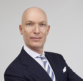
Peter Csiki
HungaryTitle: Aesthetic driven orthodontics with the Pitts Protocols
Abstract:
During my lecture I will talk about the paradigm shift with the Pitts Protocols in other words how we can change our orthodontic methods to achieve greater results with aesthetic driven treatment planning and how we can accomplish perfect aesthetics with great function. The Pitts Protocols include the super precise Pitts21 bracket system, the Smile Arc Protection (SAP) bracket positioning, early torque control, the use of disarticulation and early light elastics with a simplified archwire sequence. With the Pitts Active early concept we can reduce the treatment time by engaging and activating the appliance as early as possible and the expansion with light forces helps for non-extraction treatment to be able to create the “12 tooth smile”. You will see how we can use Digital Smile Design, tooth recontouring, interproximal reduction and laser gingiva contouring for superior esthetic results. Case Studies will demonstrate how is it possible to simplify and shorten the treatment time of the various malocclusions and how to turn extraction cases into non-extraction and surgical cases into non-surgical solution with beautiful aesthetic results.
Biography:
Péter Csiki DDS, MSc was graduated in 1994 at the Semmelweis Medical University of Budapest Faculty of Dentistry. He took his orthodontic specialty in 2000. From 1994 to 2002 he worked at the Semmelweis Medical University of Budapest Department of Orthodontics and Pedodontics as professor’s assistant. He practices in Budapest (Hungary) and in Milan (Italy). In 2004 he started to work with passive self ligation, he’s been teaching as PSL speaker since 2008. He’s also an expert in TAD, lingual braces and aligner treatment. He is an honorary associate professor at the University of Szeged Faculty of Dentistry. He is an Ortho Evolve and Ortho Classic instructor, member of the Pitts Progressive Study Group and took his Master degree in 2014 after taking part in the Pitts Masters Continuum program.
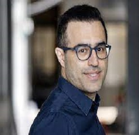
Title: Disicion-Making Non-surgical retreatment, epicoectomy, or observation?
Abstract:
Making an accurate treatment plan would be sometimes challenging for both general dentist and some specialists. When a patient refers with a root canal treated tooth that requires coronal reconstruction, the condition of the previous root canal treatment would be conclusive. Deciding about doing or refusing the retreatment prior to crown build-up is a controversial issue among many practitioners.
This one-hour presentation aims to clarify the guidelines for general dentists and endodontists about how to decide when they want to make a prosthetic treatment on previous root-treated tooth. Moreover, there would be introduced a guideline when an orthograde retreatment or retrograde surgery should be considered, when the previous root canal treatment is inadequate. What are our chances and risk factors to make a good prognose out of the previously poor treated tooth?
The presentation is based on the various literature review and guidelines from AAE (American Association of Endodontics) and E-S-E (European Society of Endodontology) as the basic of our science in decision making. Furthermore, there would be presented some clinical cases and follow-ups, managed by author, which would be simplified the understanding of the concept and will contribute to give a bright grasp for deciding on the most appropriate treatment.
Key points:
• When should a practitioner carry out a non-surgical retreatment?
• When should a practitioner carry out a surgical retreatment?
• When should a practitioner choose to monitor a previous root canal treated tooth?
• Illustrate a clear guideline for introducing an optimal treatment plan.
Biography:
Younes Alipanah has completed his general dentistry at the age of 23 years from Shiraz University and Master of science in endodontics from Shahid Beheshti Dental School, Tehran, Iran at the age of 30. Recently, Younes has received the second MSc in Endodontic from King’s College in London. He is the clinical instructor at Copenhagen dental school, and work as endodontist and referral dentist in two Private clinics.
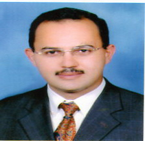
Nasser Al Alami
UAETitle: Case Report : Pansinusitis and periorbital cellulitis complicating External sinus lifting procedures
Abstract:
Perforation of the maxillary sinus membrane is a common complication during sinus lifting procedures(10-40%).Maxillary sinusitis might result and displacement of the graft material may be a contributing factor. Antibiotic Regimes with removal of the implants perforating the sinus have been used for management of sinusitis with immediate or late replacement of the dental implants .
Patient with recurrent severe maxillary,ethmoid and frontal sinusitis attended our center complaining from periorbiral cellulitis .She visited multiple ENT clinics for several months with limited improvement and recurrence of the infection.CT scan revealed titanium membrane displacement into the maxillary sinus. Drainage was done intraorally and removal of the titanium membrane. She had sinus lifting procedure right side 9 years ago for placement of 3 implants to replace upper posterior teeth. Patient insisted not to remove the implants and accepted treatment without warrranty of the result. Sinus membrane was elevared and PRF used to fill the space above the implant apices. The infection resolved completely within 10 days . Follow up of the patient for more than 40 months did not show any recurrence . CT Scan shows formation of bony partition between the implants and the maxillary sinus .
Biography:
Nasser Al Alami is a Maxillofacial Surgeon and he completed his Master of Science in Oral and Maxillofacial Surgery (2003) in Baghdad University. Nasser Al Alami Ministry Of Health and Prevention UAE

Nasser Al Alami
UAETitle: Autogenous tooth transplantation
Abstract:
Perforation of the maxillary sinus membrane is a common complication during sinus lifting procedures(10-40%).Maxillary sinusitis might result and displacement of the graft material may be a contributing factor. Antibiotic Regimes with removal of the implants perforating the sinus have been used for management of sinusitis with immediate or late replacement of the dental implants .
Patient with recurrent severe maxillary,ethmoid and frontal sinusitis attended our center complaining from periorbiral cellulitis .She visited multiple ENT clinics for several months with limited improvement and recurrence of the infection.CT scan revealed titanium membrane displacement into the maxillary sinus. Drainage was done intraorally and removal of the titanium membrane. She had sinus lifting procedure right side 9 years ago for placement of 3 implants to replace upper posterior teeth. Patient insisted not to remove the implants and accepted treatment without warrranty of the result. Sinus membrane was elevared and PRF used to fill the space above the implant apices. The infection resolved completely within 10 days . Follow up of the patient for more than 40 months did not show any recurrence . CT Scan shows formation of bony partition between the implants and the maxillary sinus .
Biography:
Nasser Al Alami is a Maxillofacial Surgeon and he completed his Master of Science in Oral and Maxillofacial Surgery (2003) in Baghdad University. Nasser Al Alami Ministry Of Health and Prevention UAE
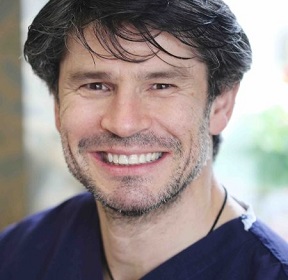
Viorel Matei
IrelandTitle: The effect of the gingival biotype on the crestal bone stability surrounding dental implants. A literature review
Abstract:
Background
Traditionally crestal bone loss has been investigated in the literature by assessing multiple variables that may have an effect on the peri-implant bone remodelling. Thus, polished implant neck, immediate loading versus late loading, and occlusion have been assessed in respect with crestal bone loss around implants. Nowadays, soft tissue biotype has been investigated as one of the factors that may influence the crestal bone stability. The objective of this study was to investigate in which measure soft tissue thickness has an influence in crestal bone loss around dental implants at 1-year follow-up. The question to be answered in this study was the following: “Is there any effect of soft tissue thickness on crestal bone stability around dental implants?”.
Methods
Electronic and manual literature search was carried out in three databases including MEDLINE, EMBASE both via OVID, and Cochrane Oral Health Group Trials Register from 1974 up to June 2020. Finally, nine articles met the inclusion criteria and were analysed in the current review.
Results
Seven of nine studies reported the differences of the crestal bone loss in thin versus thick gingival biotype, and two studies reported only the results of the crestal bone loss in thin gingival biotype. However, those two studies were included in this review due to the fact that the reported results were
in concordance with the results reported in the other seven trials included in the current review. All authors reported that thick gingival biotype shows less crestal bone loss when compared to thin gingival biotype, and a minimum of 2 mm gingival thickness is required to minimise the crestal bone loss. Furthermore, augmentation of thin biotype should be considered as an adjuvant in crestal bone preservation.
Conclusion
All studies included were at high or moderate risk of bias. Consequently, well-designed randomised controlled clinical trials are needed to accurately assess and analyse the effect of gingival biotype thickness on the clinical outcomes of peri-implant crestal bone loss.
Biography:
Viorel Matei graduated in Dentistry from Trinity College Dublin, Ireland and has a Master of Science - Implantology from the University of Manchester, United Kingdom. His master thesis focused on “The effect of gingival biotype on crestal bone stability around dental implants”. In 2014, Matei won the Leo Heslin Memorial Medal awarded by the Royal College of Surgeons in Ireland, and during his undergraduate studies he won the best prosthodontics case presentation. Throughout his professional career, he has attended numerous world-class courses and conferences to keep abreast of new techniques and technologies in dentistry. He possesses a wide range of dental experiences, particularly in the field of implant fixed and removable prostheses. Matei is a member of several dental societies, including in Ireland, United Kingdom, Netherlands and Romania, as well as the Royal Academy of Medicine in Ireland.
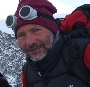
Sergei Mikhailovich Aizikovich
Russian FederationTitle: Mathematical modelling for construction of optimized biocompatible coatings
Abstract:
Statement of the Problem: Deposition of thin films and coatings on surfaces of dental implants and surgical tools can significantly change their physical and mechanical properties. Thus wear and erosion resistance, biocompatibility, durability, protection against abrasion and corrosion, thermal and chemical stability can be significantly increased. For practical researches of the mechanical characteristics of coatings both in laboratory conditions and for industrial needs, the nanoindentation technique is frequently used. This technique can be represented by a set of methods that use local precision force action on the material and simultaneously record the deformation responses with nanometer resolution. Most modern nanoindentation systems are equipped with software that allows operators to interpret experimental results. This software is built upon the methods based on the mathematical models that use solutions of classical contact problems for isotropic homogeneous materials. Such methods give the possibility to evaluate elastic moduli of bulk materials. In some cases, with their help the elastic properties of coatings can be determined. For this purpose, it is recommended to carry out indentation with depths not exceeding 10% of the coating thickness, and some specific ratios of the mechanical properties of the coating and the substrate must be fulfilled. However, these methods may radically underestimate or overestimate the unknown values of the characteristics in case of a sufficiently large difference between the elastic moduli of the coating and the substrate. It should also be taken into account that during the investigation of thin coatings, the recommended indentation depth may turn out to be comparable with the height of defects or roughness. This possibility entails high measurement errors or even an inability to conduct the experiment.To study the properties of inhomogeneous coatings required in modern dentistry one has to resort to the methods based on mathematical models using solutions of contact problems for solids with coatings. Methodology & Theoretical Orientation: In the present study, an effective mathematical model, taking into account the layered or continuously inhomogeneous structure of the sample, is proposed for the description of nanoindentation experiment. The analytical solution was obtained using the bilateral asymptotic method. A comparison of the experimental results with the results, obtained using the mathematical model was performed for ZrN (Figure 1) an ZnO coatings on various substrates. Conclusion & Significance: A good agreement was demonstrated for the results of mathematical modelling and nanoindentation test data for various coatings.
Biography:
Sergei Aizikovich, PhD, Head of Laboratory for Functionally Graded and Composite Materials of Don State Technical University, has over 110 publications in Scopus database (the publication H-index is 16), completed his habilitation (doctoral thesis) entitled “Contact problems of elasticity for non-homogeneous media” in 2003. The main research interests of Aizikovich include biocompatible coatings, functionally-graded materials, composites, nanoindentation, mathematical modelling, contact problems, biomechanics of oral cavity. The team lead by Aizikovich is currently conducting research projects on biomechanics of the tissues of oral cavity and eyeball and optimized biocompatible materials for implantation.
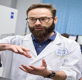
Evgenii Valerievich Sadyrin
Russian FederationTitle: Deformation behavior and biomechanical properties of enamel and dentine about a white spot lesion
Abstract:
Statement of the Problem: The appearance of acids in the oral cavity may occur during the consumption of a number of foods and beverages, and also as a result of the vital activity of cariogenic bacteria, which contributes to the demineralization process - partial dissolution of the main structural elements of enamel - hydroxyapatite crystallites. Early caries is characterized by the process of demineralization of tooth enamel without cavitation under a relatively intact tissue surface. In this case, pores appear in the subsurface region. Due to the significant difference in the refractive indices of the medium within the porous area and the sound tissues surrounding it, a whitish opaque region appearance of demineralized pathologically altered enamel lesions can be observed. This phenomenon is called white spot lesion (WSL). Overall, the number of studies characterizing the fundamental changes that emerge in natural WSLs from materials, microstructural and biomechanical point of view remains rather low. Methodology & Theoretical Orientation: The present work aims to characterize the complex of properties (mineral density, reduced Young’s modulus, indentation hardness, average roughness, maximum height of roughness) and features (surface structure, molecular composition, indentation creep) of the enamel and dentine about a WSL on a single tooth using computed X-ray microtomography, bitewing X-ray, nanoindentation, Raman spectroscopy, scanning electron (Figure 1), atomic force and optical microscopy. The aim of obtaining such information is to provide the basis for future studies of efficacy of minimal invasive treatments of caries and deeper analysis of these properties and features. Conclusion & Significance: The work reveals some fundamental changes emerging inside human enamel and dentine at the first clinically visible stage of the carious disease from several points of view. The complex of characteristics was found for the natural enamel WSL and dentine bordering it. The properties obtained were compared to those of sound counterparts of the aforementioned areas on the same tooth. The significant reduction of the mechanical properties was recorded for both carious areas accompanied by the abnormality of the unloading response and change of the character of indentation marks caused by mineral loss. Increase of indentation creep for WSL area was recorded. The maps of mechanical properties show that the enamel outside the WSL is weakened despite its appearance as being sound on optical and bitewing X-ray images. At the same time, the decrease in mineral density of the WSL area and bordering dentine was rather small. The average roughness of polished surfaces was considerably increased in the caries-affected areas caused by the demineralization process, and the surface relief changed as well. Raman spectroscopy research resulted in detection of interesting repeatable features connected to changes in the molecular composition in the carious areas. This research was funded by the Russian Science Foundation, grant number 19-19-00444.
Biography:
Evgenii Sadyrin, M.S., Junior Researcher of the Laboratory for Mechanics of Biomaterials, Don State Technical University, is the author of 40 publications in Scopus database (the publication H-index is 8). The main research interests of Sadyrin include biomechanics of tooth tissues, nanoindentation, microtomography, atomic force microscopy, scanning electron microscopy, Raman spectroscopy, biocompatible coatings, mathematical modelling. Sadyrin is currently involved in research projects on biomechanics of the tissues of oral cavity and eyeball and optimized biocompatible materials for implantation.
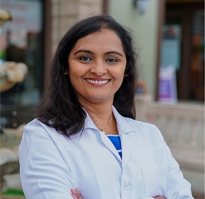
Savitha Siddappa
USATitle: An overview of Temporomandibular disorders etiology, diagnosis and management in your practice
Abstract:
Temporomandibular disorders (TMD) is a collective term embracing a number of clinical problems that involve the masticatory musculature, the temporomandibular joint (TMJ) and associated structures, or both" (De Leeuw, 2008. Basically, musculoskeletal pain disorders of the masticatory system.
In this presentation, I will give you an overview of TMJ disorders, etiology, signs and symptoms, options available in the management and make decision whether to treat or refer in a general practice. The diagnosis goes beyond jaw pain, clicking and popping of the jaw. Management is not just giving the patient a nightguard and hope for the resolution the issue.
Biography:
Savitha Siddappa received her DMD degree from Henry M Goldman School of Dental Medicine, Boston MA followed by an AEGD from Lutheran Medical Center, New York, MS in Orofacial Pain and Oral medicine from University of Southern California, Los Angeles. Currently practicing as a orofacial pain specialist in greater Los Angeles area. In the last 2 decades of being in dental field, she has invested a lot of time into learning the latest advances in various aspects of managing different dental diseases. She enjoys learning new technologies in the field of dentistry and have implemented it in her practice throughout her career. Outside of work, she enjoys hiking, travelling and spend time with her family.
She is a Diplomate, American Board of Orofacial Pain Fellow; American Board of Orofacial Pain Fellow; American Academy of Oral medicine; Member of mentorship committee of AAOP
Zuzanna Grzech-Leśniak
PolandTitle: Periodontitis management with laser therapy as a new treatment option
Abstract:
Statement of the problem: Periodontitis is a disease that affects the whole organism, leading to cardiovascular diseases, respiratory disorder, chronic renal disease, diabetes type 2 and many others. The origin of this condition is supra-or subgingival bacterial biofilm that leads eventually to the destruction of the periodontium. Not that far ago the only way to counteract the discussing disorder was mechanical therapy with additional antimicrobial and local or systemic antibiotic therapy. Increasing antibiotic resistance has led to rise to a new modern treatment approach. During oral presentation will be presented result of using periodontitis treatment with high power lasers. Lasers clinically can reduce risk of side effects to minimum. Laser offers a lot of advantages such as selective calculus ablations, bleeding control, bactericidal and detoxification effects. The purpose of the lecture is to illuminate the current role of lasers in periodontal treatment procedures. The use of laser technology is becoming more and more popular in everyday dental practice, since it provides better treatment outcomes compared to conventional methods. The aim of the presentation is introduce the studies on our research group which were focus on the evaluation of clinical
and microbiological findings following non-surgical periodontal therapy by the different following
methods: traditional periodontal debridement with a combination of ultrasound and hand curettes; antimicrobial photoactive disinfection with diode laser, combination of neodymium and erbium lasers high-power diode laser with combination of oxidative disinfectant as hydrogen peroxide or sodium hypochlorite
Findings: All therapy modalities resulted in the superior clinical effect comparison to traditional mechanotherapy including reduction of total bacteria count as well as red and orange complexes according Socranskyetal. Clinical research results show that after laser treatment therapy there is significant reduction of plaque index as well as bleeding on probing with the reduction of periodontal probing depth. The best combination that gives the most promising outcomes is combination diode laser with root scaling and low concentration of sodium hypochlorite, hydrogen peroxide or combination of two high power lasers like neodymium and erbium lasers in one procedure.
Conclusion: Research findings constitute beneficial alternative laser assisted non-surgical periodontal treatments in comparison to conventional treatment.
Biography:
Zuzanna Grzech-Leśniak is a young passionate scientist that is specially interested in innovative new approaches to dental treatment. Her special field of interest is laser therapy. She is author of international and national publications. She is associated with the Wroclaw Medical University that is at the forefront of Polish medical universities that are present in the prestige international academic rankings, such as Shanghai List or Best Global Universities.
Speakers
Gaurav Vishal
IndiaTitle: Squamous Cell Carcinoma of the Buccal Mucosa: Staging and Surgical Management
Abstract:
Introduction: Squamous cell carcinoma of the buccal mucosa is the most common oral cavity cancer in Southeast Asia. In India, 60 to 80% of oral squamous cell carcinoma cases present with advanced stage as compared to 40% in developed countries. Carcinoma of the buccal mucosa is treated mainly by surgery followed by adjuvant therapy, depending upon the stage (early and advanced) and histopathological characteristics. The purpose of this study was to evaluate the staging (AJCC 8th edition) and surgical management of squamous cell carcinoma of the buccal mucosa.
Methodology: A total of 54 histopathologically proven cases of oral squamous cell carcinoma of buccal mucosa who had no previous malignancies were included in our study. Recurrent cases and prior treatment of oral cancer by chemotherapy and radiotherapy were excluded. All the patients involved in the study underwent tumor resection with neck dissection.
Results: All 54 (46 male and 8 female) cases were staged as per TNM criteria (AJCC 8th edition). The percentage of T1, T2, T3 and T4 lesions were 5.56, 27.78, 12.96 and 53.7% respectively. Twenty four cases (44.44%) presented in N0 stage, 10 (18.52%) in N1 stage, 9 (16.67%) in N2 stage and 11 cases (20.37%) presented in N3 stage. The lymph node positivity was highest in T4 followed by T3 and T2. Final histopathologic stage grouping revealed stage I in 3 (5.56%) patients, stage II in 8 (14.81%) patients, stage III in 8 (14.81%) patients, and stage IV in 35 (64.82%) patients. 9, 34 and 11 patients were treated by surgery alone, surgery with postoperative radiotherapy and surgery with postoperative CTRT respectively.
Conclusion: Majority (79.63%) of the patients had diagnosed in advanced stage (stage III and stage IV) of carcinoma. One-fifth (20.37%) of the patients were pathologically node-positive with extranodal extension (pN+/ENE+). Histopathology reports revealed the most of the cases had well-differentiated squamous cell carcinoma of the buccal mucosa. Early stage (stage I and II) patients were treated primarily by surgery alone and advanced stage (stage III and IV) patients were treated with combination therapy.
Biography:
Gaurav Vishal is an Oral and Maxillofacial Surgeon (M.D.S), Fellow in Oral Oncology and Reconstructive Surgery. He completed M.D.S- Oral and Maxillofacial Surgery from Institute of Dental Sciences, Bareilly International University, Bareilly, India in 2020 and Fellowship in Oral Oncology and Reconstructive Surgery from Rohilkhand Medical College and hospital, Bareilly, India in 2021 under the guidance of Dr. Arjun Agarwal (Surgical Oncologist) and Dr. Anurag Yadav (Oral, Onco & Reconstructive Surgeon).
Aamna Rahat
IndiaTitle: Behavior Changes in Children of Different Age Groups During Dental Appointment with or without Local Anesthesia by using Modified Venham Behavior Rating Scale
Abstract:
INTRODUCTION: The basics of practicing child dentistry is dependent on the potential to guide them through their dental experiences. The idea of treating the patient and not just the tooth should be operative with all patients but it is more important with pediatric patients. The prime aspect of child management in the dental chair is managing dental anxiety, a world-wide problem and a universal barrier to oral health care.
AIM: This study was planned to assess the behavior changes in children during procedures with and without local anesthesia by using modified venham behavior rating scale.
MATERIAL & METHOD: It consists of 101 children of age 3-14 years and they further divided into 2 sub-groups i.e. 3-8 years & 9-14 years. Children required a procedure with LA (2% lignocaine with 1:80,000 adrenaline) and should have 1 previous & 1 next dental appointment in which procedures without LA need to be performed, were included in the study.
RESULT: Total cooperation was least during 1st visit (procedures without LA) than the 2nd visit (procedures with LA) & it was maximum during 3rd visit (procedures without LA). Hence the prior experiences could influence the behavior of children at subsequent dental visits. There was a significant difference between 3-8 years and 9-14 years old with respect to behavior at 2nd and 3rd visit, although there is no significant difference in the 1st visit.
CONCLUSION: Fear and anxiety influence child’s behavior to a greater extent which establishes the success of dental treatment, it is foremost to identify the anxious children at the initial age possible inspite to deal with them later, consecutive dental visits have been shown positive effect on psychological response that causes the improvement in behavior even during the procedures including local anesthesia. Therefore, the influence of 1st dental visit can affect all future reactions and behavior to the dentistry.
Biography:
Aamna Rahat is a pediatric dental surgeon, working as a Assistant Professor in the Department of Dentistry at SRMS Institute of Medical Sciences, Bareilly, India. She has completed her “Master of Dental Surgery” in the subject of Pedodontics And Preventive Dentistry from Bareilly International University. She is University Gold Medalist for M.D.S and done her B.D.S from Teerthanker Mahaveer University, Moradabad, India. She has Received Medal for her B.D.S Program too. She has several paper publications and international E-book publications.
Dania Wail Islam
Saudi ArabiaTitle: Molar Incisor hypomenaralisation (MIH)
Abstract:
The new abbreviation of molar incisor hypomineralisation, MIH, was first launched in 2001. Increasing attention has been paid to this pediatric dental problem worldwide along with improving level of children’s dental health. Since 2001, a total of 96 original articles have been published on children’s molar incisor hypomineralisation using the MIH-abbreviation in the title or in the abstract. The main clinical characteristics of MIH-lesions are demarcated brownish or chalky white opacities of enamel, atypical restorations, post-eruptive breakdown of enamel, and extractions due to MIH. Microscopically, the opacities reveal reduced microhardness, reduced mineral content, large areas with porosities, and defective enamel rod and surface ultrastructure. Some of the MIH-molars are highly sensitive to tooth-brushing and instrumentation. Moreover, children with severe MIH-lesions are at risk of developing severe dental fear and anxiety due to repeated exposure to restorative and operative dental care. MIH-lesions are most common among first permanent molars and incisors, but are also seen in deciduous second molars. The overall prevalence of MIH is about 13-14%, ranging between 2.8 and 21.8%. The problem seems to be fairly common not only in Europe but in Australia, New Zealand, India, China and South America as well. In spite of extensive research and publications, the etiology of MIH-lesions is far from clear. Several reasons like prematurity, gastrointestinal problems, pneumonia, frequent fever episodes, measles, chicken pox, bronchitis, hypoxia, hypocalcemia, and exposure to antibiotics and dioxin have been linked to MIH-lesions. Due to the still unknown etiology, all levels of prevention should be exercised and combined with meticulous restorative dental care of these pediatric patients. Local anesthesia may be problematic in hypersensitive MIH-molars, and adhesive techniques in restorative care of severe MIH-lesions can be challenging due to the defective ultrastructure of enamel. Moreover, extending borders of the restoration to areas where no further risk of tissue breakdown exist can also be challenging.
Biography:
Dania Wail Islam did the DDS.MS.Diplomat in the American Board of Pediatric Dentistry and she is a consultant in pediatric dentistry & early orthodontics. She is the assistant professor at university of Malaya in Saudi Arabia and she is married and a mother of a wonderful daughter
Poster
Alisa Vladimirovna Blinova
Russian FederationTitle: Antibacterial effect of the new intracanal medication based on copper-calcium hydroxide and silver nanoparticles: real-time PCR study
Abstract:
Alveolar bone regeneration may be achieved only in 53.6% - 70.8% of apical periodontitis cases. The root canal system has an irregular shape, which is difficult to clean with endodontic instruments. Besides, zones of exposed root dentin, formed during root canal cleaning, include a large surface area – there are 20,000-80,000 dentine tubules’ apertures on each 1 mm2. Persistent bacterial biofilm is found in dentine at the depth of 300 – 1000 microns. So, it’s necessary to develop new ways of antimicrobial treatment. The aim of the study was to evaluate the antibacterial properties of a new intracanal paste based on calcium hydroxocuprate (CHC, Ca[Cu(OH)4]) and silver nanoparticles hydrosol for root dentine impregnation. The study included 55 teeth with 69 root canals of 29 patients with chronic apical periodontitis. The study did not include patients with decompensated chronic somatic disorders and acute infectious diseases, as well as patients with obliterated root canals or allergic reactions to copper and silver medicines. Informed consent was obtained. First, the tooth was isolated with a rubber dam. After cavity preparation, the root canals were minimally expanded. The first biological sample was taken with a paper absorber. The canals were mechanically treated and irrigated with a 3% solution of sodium hypochlorite. Then the canals were filled with antimicrobial paste. The main group, including 44 root canals, was filled with a new paste based on CHC and silver nanoparticles for 7 days. CHC paste «Kupral®» (Humanchemie GmbH, Germany) and a colloidal solution of silver nanoparticles (3 mg/l, particle size is 0,5 - 3 nm) were mixed in a ratio of 1:1. Silver hydrosol was obtained by condensation of low-temperature plasma in a spark discharge. In the control group, 25 root canals were sealed with an aqueous paste of calcium hydroxide «UltraCal®» («Ultradent», USA) for 14 days. During the second visit, the paste was washed out of the root canals with a sterile isotonic solution and the second biological sample was taken. The presence of the endodontic microorganisms in each sample was evaluated by real-time PCR. Statistical analysis was performed using SPSS software. Significance levels were set at the 5% level using the Student t-test or Mann-Whitney test. 95% confidence intervals (CI) were calculated.
At the first step, the comparability of the groups was checked. No significant differences in the bacteria species were found between the groups before treatment. Further analysis showed that the amount of the DNA, common for P. gingivalis, T. forsythia and T. denticola, after treatment was less in the main group, where the new paste was applied. These results were significant at the p = 0.05 level (p=0,005, p = 0,006, p=0,003 according to each mentioned bacterial species). No significant differences were found between the groups in the number of genome equivalents specific for P. intermedia and
F. nucleatum (p=0,543, p=0,554), although the amount of DNA of these bacteria in the main group was less than in the control group. These findings suggest that the new method of root dentine impregnation with the CHC and silver nanoparticles paste may be useful for the treatment of chronical apical periodontitis. The new medication turned out to be more effective than calcium hydroxide against the most pathogenic microorganisms.
Biography:
Alisa Vladimirovna Blinova, PhD, a post-graduate student of Tver State Medical University (Russia). She graduated from Tver State Medical University in 2020 with a degree in Dentistry. She has over 40 publications and 5 patents that have been cited over 30 times (the publication H-index is 3). Her research interests include modern conceptions of endodontic treatment, the control of oral biofilms and using nanotechnologies in dentistry. The method of passive nano-impregnation developed by her has a high therapeutic and commercial potential.
Speakers
Abdelrahman Nasiem Ahmed
EgyptTitle: The effects of the face mask on soft tissue profile with or without expansion A Systematic Review
Abstract:
Background
Face mask treatment on growing patient produce skeletal and dental effect and different studies stated that face mask produce improving effect on soft tissue profile that following under skeletal profile changes and other studies were stated that no significant effect of face mask on soft tissue so no evidence found on literature on effect of facemask on soft tissue profile with or without expansion used with face mask for maxillary protraction treatment.
Objective
Main objective in this review to evaluate evidence of effect of Face mask in soft tissue profile in treatment of skeletal class Ill growing patients with or without maxillary expansion.
Search Strategy
Following key words were used in different electronic database search Facemask-skeletal class maxillary expansion - maxillary protraction- soft tissue profile-reverse pull head gear detailed search strategy were performed in following electronic database (Pumped-Google scholar- Cochrane electronic data bases) to collect all eligible studies in English language that matching inclusion criteria of this review.
Selection criteria
The following criteria were met inclusion criteria for study selection: controlled or non controlled clinical trials on growing patients receiving orthopedic face mask treatment with or without maxillary expansion or retrospective studies with soft tissue profile measurements before and after treatment. Studies that were involved adult patient or fixed appliance combined with face mask treatment or surgical treatment or cleft patient or any other dento facial defects were excluded also Studies.
Biography:
Abdel-Rahman Nassim received his BD in 2009 from October 6 University and his orthodontic MD in 2016 from Cairo University. From 2016 till now he carried the burden of educating patients about the importance of orthodontic treatment and training dentists to orthodontic basics that every dentist should know in orthodontics. In 2020, Nassim was the first dentist who began studying the sterilization process and comparing between class B, Class N and high-pressure steam sterilizer autoclave
Title: Non-extraction Treatment of Adult Class II Subdivision Malocclusion; Two Different Techniques
Abstract:
Treatment of adult Class II subdivision malocclusions can be challenging for clinicians because it is inherently difficult to treat asymmetric malocclusions than symmetric ones. Class II subdivisions can be addressed using different approaches including asymmetric extractions, orthodontic distalizers, fixed functional appliances and surgical orthodontic therapy. The choice of treatment approach depends on the cause of asymmetry, patient’s age, skeletal pattern and patient’s profile. Two cases will be presented to illustrate different techniques for correcting class II subdivision malocclusion in adult patients. Both patients have class II division 2 subdivision. The first case was treated with lingual fixed appliances and forsus device, and the second case malocclusion was corrected with labial fixed appliances and sliding jig. The appliance design, treatment sequence and treatment outcome of each case will be presented.
Biography:
Fadwa A Shembesh, an American Board Certified Orthodontist. She is currently an orthodontic faculty member in Benghazi University and Orthodontic Specialist in Jana Dental Center. She received dental degree (Bachelor of Dental Surgery BDS) in 2008 from Benghazi University, Faculty of Dentistry, where she graduated with honors as the first of the batch. Because of her passion for Orthodontics, she moved to the United States to finish her postgraduate studies and pursue the dream of becoming an orthodontist. Her journey in the States started with a one year Advanced Clinical program for International dentists in Orthodontics at New York University, College of Dentistry (NYUCD). Then the journey continued by finishing three year CODA accredited Orthodontic Residency Program at New York University in 2017. She became Board Certified Orthodontist in September of the same year and received T.M Graber outstanding article award in 2019. She is an active member of the American Association of Orthodontists and North Eastern Society of Orthodontists. She has active dental licenses to practice in the States of Texas and Oregon.
Abdulaziz Abdulhadi
United Arab EmiratesTitle: Evaluation of the functional treatment of skeletal Class II malocclusion with low-level laser-therapy-assisted Twin Block appliance: a three-arm randomized controlled trial
Abstract:
Background: Different techniques have been used to reduce functional treatment time including low-level laser therapy (LLLT) and the majority of studies have been conducted on animals. Therefore, the aim of the current study was to evaluate the effects LLLT on improving orthodontic functional treatment.
Methods: Study design: This study was three-arm, parallel-group, randomized controlled trial.
Patients were selected using the following inclusion criteria: skeletal Class II division 1 malocclusion (SNB angle 73-78°), ANB angle between 4-9°, over-jet between 5-9 mm. Forty-eight patients were randomly allocated into three equal groups. In the LLLT-TB group, the low-level laser device was used with a wave length of 808 nm and power of 250 mWatt in addition to functional treatment with Twin block appliance. The laser was applied on the skin at the bilateral temporomandibular joint (TMJ) regions, at five points, each point received 5 joules of laser during 20 seconds. The laser course was twice a week in the first month, and every two weeks in the second month, and every 3 weeks up to the end of treatment. The TB group received functional treatment with Twin block appliance, while the control group had no treatment for 9 months.
Results: There were statistically significant differences in treatment periods between the LLLT-TB group and the TB group (129 days, 235 days respectively, p-value <0.001). The change in the effective mandibular length (CoGn) was the highest in the LLLT-TB group compared with the TB and control groups (4.41 mm, 3.66 mm and 1.07 mm respectively, P-value <0.001).
Conclusions: Application of low-level laser on the condylar region accelerates functional treatment and linear measurements were higher in laser group than in the control one.
Registration: This trial was registered at Clinical Trials.gov with a TRN (NCT03239912) on August 4, 2017, before the enrollment of the first participant.
Keywords: Functional treatment, Class II malocclusion, low-level laser therapy
Biography:
Experienced Orthodontist with a demonstrated history of working in the higher education industry. Skilled in Strategic Planning, Research, and Management. Strong healthcare services professional.

