Speakers
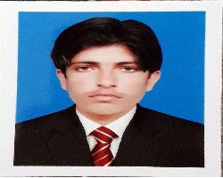
Dr Mahesh Kumar
Graphic Era Deemed to be University IndiaTitle: Mems impedance flow cytometry: A computation analysis for biological cell motion manipulation
Abstract:
Efficient manipulation of microbiological cells has always been an important task in the healthcare sector. For this purpose, dielectrophoresis (DEP) based microfluidic cell separation devices have been proven to be a promising technology. The DEP technique uses the dielectric properties of the micro-entities such as micro-particles and biological cells to manipulate the motion in the fluidic medium. The motion manipulation of cells/microparticles can be achieved using a non-uniform electric field in the microchannel with an appropriate external AC supply. The DEP forces generated in the micro-channel depend upon various biological and physical parameters of cell and suspending medium. Apart from that, design parameters of the microfluidic channel and dimension of electrodes used for generating DEP action also play a major role in microcell/bead manipulation. This session will cover the computational analysis approach on microfluidic device optimization and future perspectives for efficient biological cells separation by interrogating electrode structure, channel dimensions, and other electrical aspects in the devices with the finite element method.
Biography:
Mahesh Kumar recently completed his PhD research at the age of 32 years from Manipal University Jaipur (India) on topic ‘A Microfluidic Device for Efficient Diepectrophoretic Separation and Sensing of Biological Cells. He ha salso completed his masters from IIT Kharagpur (India) in the area of microfluidics. He is currently working as Assistant Professor in Department of Computer Science and Engineering, Graphic Era Deemed to be University Dehradun, India. He has published several research papers in reputed journals and has been served in many reputed journals as journal reviewer.
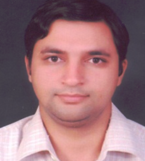
Dr Manish Gupta
University of Pennsylvania, USA United StatesTitle: Development of Potable and Affordable Nuclear Magnetic Resonance (NMR) for Malaria Diagnosis in Human Subject
Abstract:
Malaria is a leading parasitic disease which imperil the lives of many people especially in tropical climates. Presently, available malaria diagnostic tools/ techniques e.g. [Giemsa-Stained Microscopic, Rapid Diagnostic Tests (RDT), and Polymerase Chain Reaction (PCR) are less sensitive, time consuming, requires well trained operator, and costly in nature. In additions, both PCR and RDT are unable to provide quantitative analysis on the parasitaemia levels. Hence, there is an urgent demand of fast, sensitive, and reliable diagnostic tools. The portable Nuclear Magnetic Resonance (NMR) has been gaining considerable attention amongst scientist community due to the promising applications in in-vivo and quantitative detection of various small biological/chemical applications related to healthcare. Traditionally, available NMR is very costly, requires skill operator, bulky and thus difficult for Point- of-Care applications. Here, in this talk a portable, cost-effective, and benchtop NMR system having 0.5 Tesla field operating at 21.287MHz is demonstrated. The developed NMR is successfully capable of detection of the parasite load during the early stage of Malaria.
Biography:
Dr. Manish Gupta is currently a postdoctoral researcher at Department of Radiology, University of Pennsylvania, USA. He completed his Ph.D. from Ph.D. in Computer Science from JNU, New Delhi. Dr Gupta is also former postdoctoral researcher at Gwangju Institute of Science and Technology, South Korea. His research areas are development of portable sensors, development of radiofrequency coils for NMR, MRI, and open magnetic particle imaging. Dr. Gupta published many research papers in reputed SCI journals with high impact factors and reputed international conferences. He also fields two patent. He is reviewers of many reputed journals including IEEE Sensors Journal. Dr. Gupta is chief research advisor of AI start-up company i.e., Arogyapandit private limited, India.
Dr Praveen kumar sunil
Saveetha Engineering College IndiaTitle: Cost-effective fabrication of porous silicon for optical biosensing applications
Abstract:
In optical biosensing technique, since the reactions are generally carried out only in close proximity to the electrode surface, the electrodes themselves play a crucial role in the performance. Here, the method for cost-effective fabrication of porous silicon (PSi) electrode is proposed, that can be used as a biosensor substrate due to its large surface area, good refractive index, high biocompatibility, and reproducibilty without any surface modification. If resistivity of silicon wafer is increased, the wafer cost is decreased. Hence, the high resistive p-type silicon wafer is used here to fabricate the PSi. Electrochemical etching process is used to synthesize the PSi from the high resistive bulk silicon. The etching parameters like current density is maintained as constant as 10 mA/cm2 and etching time are varied as 60, 120 and 180 min. Electrochemical etchant like HF and isopropanol are mixed in the ratio of 1:2. The porosity is determined by gravimetric method and the photoluminescence and spectrum absorption are investigated by using the spectroflurometer. The formed PSi surface morphology is analyzed using SEM study. The obtained photoluminescence response shows that if the porosity is increased, then the band gap is increased. The maximum porosity achieved is 84.21% for the etching time 180 mins and maximum energy band gap is obtained as 1.8921 eV for the wavelength 655.35 nm with refractive index of 2.874. The results revealed that the fabricated PSi substract has uniform pores with larger surface area and good refractive index which is needed requirement for optical biosensing applications.
Biography:
S Praveen Kumar has completed his PhD at the age of 34 years from Anna University. He is working as professor and he is the head for centre for micro nano design and fabrication, saveetha engineering college, chennai. He has published more than 15 papers in reputed journals and has been serving as an reviewer member of many reputed journals. His research area includes MEMS, BioMEMS, Microfluidics, Biosensors, Nanosynthesis, etc.
Dr Muhammad Asad
Kohat university of scince and Technology PakistanTitle: Colorimetric acetone sensor based on ionic liquid functionalized drug mediated silver nanostructures
Abstract:
The current work reports the drug-mediated synthesis of silver nanoparticles (AgNPs) and its functionalization with ionic liquid(IL) for acetonedetermination. The rationale behind the selection of Augmentin drugs was the aromaticity in its structure and the functional groups attached. Theseproperties are not only supposed to work in the synthesis of the nanoparticles but also enhance their electron density.The nanoparticles were further coated with 1-H-3-methylimidazolium acetate IL, having conductivity and aromaticity in its structure.The synthesized nanoparticleshave been characterizedby different techniques such as FTIR, XRD, SEM,and EDX, etc.Colorimetric determination of acetone was done by using IL capped AgNPswith the assistance of NaCl solution and results were analyzed by UVVis spectrophotometry. Low cost, stable eosin dye work as a substrate and is consumed resulting in a color change from brown to transparent. Different parameters such as (concentrations, loading ofnanoparticles, time and pH, etc.) were optimized to get the best results of the proposed sensor.The sensor shows awide linear range of (1×10⁻⁸-1.6×10⁻⁶M), low limit of detection 1.87×10⁻⁷Mand limit of quantification 6.22×10⁻⁷Mwith an R2value of 0.999.The proposed sensor has been successfully appliedtodiabetic patient’s urine samples for acetone detection with a visible colorimetric change. It showed good sensitivity and selectivity towards acetone detection.
Biography:
Muhammad Asad from Pakistan. He had completed his Masters degree in Chemistry with specialization in Physical Chemistry from Kohat University of Science and Technology (KUST) KP Pakistan. His research interest is in the synthesis, characterization of nanomaterials, ionic liquids, Biosensors fabrication and optimization, Paper Based Sensors and nanosensors. He have got experience in different projects and published four research papers in (Microchemical Journal https://doi.Org /10. 1016 /j microc.2020.105382) (4.84 I.F), Materials Chemistry and Physics https://doi.org/10.1016/j.matchemphys.2021.124289 (4.09 IF), Arabian Journal of Chemistry https://doi.org/10.1016/j.arabjc.2021.103164 (5.16 IF), ACS Omega https://doi.org/10.1021/acsomega.1c04548 (3.51 IF) and one paper has been accepted in Materials Chemistry and Physics (4.09 IF) hopefully at will be online at the end of this month in and two other manuscripts are under review.
Dr Divagar Murugan
Indian Institute of Technology IndiaTitle: Plasmonic fiber optic absorbance biosensor for the diagnosis of infectious diseases.
Abstract:
Detection of molecular biomarkers for the early diagnosis of infectious diseases is crucial for timely treatment and the prevention of the spread of infection. Bacterial and viral diseases such as tuberculosis and COVID-19 require faster and simpler tests. Over the past decades, optical sensing technologies have been widely explored and proven to offer sensitive, compact and affordable healthcare diagnostics. In the present work, a plasmonic fiber optic absorbance biosensor (P-FAB) for tuberculosis and COVID-19 was developed. P-FAB involves a sandwich assay, and it exploits the U-bent fiberoptic sensor platform with high evanescent wave absorbance (EWA) sensitivity, and gold nanoparticles (AuNP) labels with high optical extinction coefficient, in addition to a simple LED-photodetector based optical instrumentation. In the process of realizing a P-FAB for these applications, novel surface functionalization strategies for polymeric optical fiber (POF) by means of graphene oxide (GO) and dendrimers were also established. Optimum conditions for the realization of sensitive assays, including fiber probe geometry (fiber core and bend diameters), AuNP size, their bioconjugation, and their concentration, have been investigated in detail. Subsequently, the P-FAB for mannose-capped lipoarabinomannan (ManLAM or Mtb-LAM) and SARS-CoV-2 N-protein detection towards urine-based tuberculosis and saliva-based COVID-19 diagnosis, respectively, was established. Finally, the overall research resulted in the development of a compact point-of-care (PoC) PFAB device for diagnostic applications.
Biography:
Mr. Divagar M was born in Chennai, Tamil Nadu, India, in 1993. He received the B. Sc. Degree in Biotechnology from SRM university in 2014 and M.Sc. degree in Nanoscience and Nanotechnology from the University of Madras, Chennai, in 2016. Now, he is pursuing his PhD degree through the Department of Science and Technology (DST) - Innovation in Science Pursuit for Inspired Research (INSPIRE) research fellowship at IIT Madras, in the field of fiberoptic biosensors. He is the author of 11 research articles and 1 invention. His research interests include fiber-optics, biosensors, spectroscopy, medical diagnostics, Infectious diseases, nanomaterials, and nanostructures. Mr. Divagar M was a recipient of the Science Academies summer research fellowship award (2015), Newton-Bhabha PhD placement award (2020-21), Gandhian Young Technological Innovation award (2020), Indian Institute of Technology Madras Research Award (2021), and Advanced research opportunity program (2021-2022) - scholarship sponsored by RWTH Aachen University, Germany.
Dr Deepak kukkar
Hanyang University Republic of KoreaTitle: The expanding role of metal-organic frameworks in sensing of pollutant pesticides
Abstract:
The expanding role of metal-organic frameworks (MOFs) for the recognition of hazardous materials can be assigned to their numerous intriguing properties. Some unique characteristics of MOFs include dynamic porosity, high pore volume, tailorable optical properties, myriad of functionalities, and open metal sites. Besides, MOFs can harbor various guest molecules (e.g., metal nanoparticles, proteins, enzymes, and so on) both at their surface and within their pores. The above-mentioned properties of MOFs justify their expanding role in sensing applications. Accordingly, this work elaborates about the recent progress in the sensing applications of MOFs with specific focus on organophosphate pesticides (OPPs). Specifically, for OPPs sensing, acetylcholinesterase (AChE) was encapsulated in zeolitic imidazolate framework (ZIF)-8 to yield a nanoreactor (NR) termed as AChE@ZIF-8 NR. This NR produces a characteristic yellow color upon hydrolysis of acetylthiocholine chloride (ATCh) under optimized conditions of pH, temperature, and substrate concentration. The colorimetric biosensing of OPPs (e.g., malathion, glyphosate) was based upon their suppressive action on the ATCh hydrolysis activity of the NR. As a result, the intensity of the yellow color produced during ATCh hydrolysis by the NR is substantially reduced. Overall, the NR exhibited excellent reproducibility, stability, and ease of detection of the OPPs inhibitory colorimetric response.
Biography:
Dr. Deepak Kukkar completed his Ph.D. in 2014 from Panjab University, Chandigarh. He worked as Assistant Professor at Sri Guru Granth Sahib World University, Fatehgarh Sahib, Punjab, India from 2012 to January 2021. He was at Hanyang University as Visiting (Assistant) Professor from January 2021 to January 2022. He has published 45 articles in SCI journals. Dr. Kukkar has also served as reviewer for many Journals like JACS, ACS-Sustainable Chem and Engineering, Chemical Engineering Journal, Environmental Research, and many more. He has also served as Topic Editor for MDPI journals as well.
Dr lokendra singh
University of Engineering and Technology Roorkee IndiaTitle: Development of LSPR based Optical Fiber Sensors for the diagnosis of Biomolecules
Abstract:
Optical sensing technology is a recent and accurate measurement technology for the development of optical biosensors. Plasmonic optical sensors nowadays are employed in a plethora of applications ranging from environmental monitoring to bio (chemical) sensing. In brief, these sensors are applicable for biomedical diagnosis, drug discovery & therapies, material analysis & shaping, (bio) chemical sensing, and environmental monitoring. Plasmonics is a promising field of technology that examines the interaction between light and metallic NSs at the metal–dielectric interface. The commonly employed plasmonic-based methods such as surface plasmonic resonance (SPR) and localized SPR (LSPR). The different structures of optical fiber have been utilized to develop the biosensors. The LSPR effect in optical fiber sensors is introduced by immobilizing the layer of metallic nanostructures over the sensing regions. The mostly used metallic nanostructers are nanoparticles of gold (Au) and Silver (Ag). However, with LSPR effect the biocompatibility o optical fiber sensors is also an critical concern, which can be attained by immobilizing a layer of graphen oxide (GO) over the metallic nanostructures. The GO possess a layer of carbon bonds with very high biocompatibility for biomolecules. The development of optical fiber sensors involves various different steps such as fabrication of optical fiber structures, metallic nanostructure synthesis and their immobilization on sensing region, and functionalization of specific enzymes. The funcationaliztion of specific enzyme increases the selectivity of sensors against particular analyte. The performance analysis of optical fiber sensor structures is done by testing their ability in terms of reproducibility, reusability, sensitivity and specificity.
Biography:
Dr. Lokendra Singh has completed his PhD at the age of 29 years from DIT University and postdoctoral studies from Liaocheng University, China. Currenlty, he is the Ph.D. coordinator at University of Engineering and Technology Roorkee, India. His current research interest are optical fiber sensors, biosensors, photonic and plasmonic devices. He has published more than 40 papers in reputed journals and conferences. He has been invited as speaker in number of international conferences. Recently, he has published a noval work while introducing a plus shaped cavity in optical fiber and used for sensing application.
Dr Suryasnata Tripathy
IIIT Surat, India IndiaTitle: A Comprehensive Approach for developing low-cost portable diagnostic devices for clinical decisions
Abstract:
Healthcare is undoubtedly the most fundamental and socially alarming issue in developing countries like India and other economically challenged sections of the global community. Till today, it remains a challenge to provide easily affordable healthcare to the ones who are the most vulnerable. In particular, the unavailability of early and effective diagnostic tools is vexing, which inevitably delays clinical decision-making, leading to morbid consequences. Needless to say, there is an urgent need to address the existing shortcomings and bridge the gap between different sections of society in terms of healthcare and diagnostics. Focusing on diagnostics only, one would agree that the need of the hour is the development of next-generation sensing platforms that are capable of accurate decision making while ensuring cost-effectiveness, easy operation, and large-scale deployment. Thankfully, the recent technological advancements in device-fabrication methods and the improved understanding of nanoscale biomolecular structures and biological principles, have paved the way for the realization of highly complex and sophisticated miniaturized platforms today. Such sensors can cater to a myriad of important biological applications, including detection of DNA hybridization and antibody-antigen interactions, leading to disease diagnosis. As a next step, such sensors have to be converted into portable platforms, for desired clinical diagnosis. Such a translation involves several challenges, such as – miniaturization of the sensor, development of a portable readout and development of a data-analysis and decision-making algorithm. Addressing these challenges, while keeping the cost and the ease-ofuse in mind, is indeed an uphil task. In line with this, in this talk, considering a chemisresistive biosensing platform, a complete sensor-to-prototype pathway is described. As a part of this, the present discussion also addresses several important practical problems, namely – 1. adhesion issues at sensor interfaces, 2. inter-device variability in nanofibers, and 3. non-linearity of resistive sensors, among others. In addition, a couple of innovative sensor-fabrication approaches (such as fabless device development from commercial PCB boards and printable-bioelectronic systems) have been discussed herein, which can be adopted towards reducing the overall system-cost, without compromising with the desired performance. Further, the development of portable bioelectronic readouts has also been discussed here. Also, towards improving the overall robustness of modern diagnostic methods, a smartphone-integrated machine-learning-based decision-making process has been confabulated here. Such a process can potentially replace the conventional calibration techniques, and incorporate the benefits of AI, for rendering smart clinical decisions
Biography:
Dr. Suryasnata Tripathy has completed his Ph.D. in May 2020, in Microelectronics and VLSI, in the Department of Electrical Engineering, at the Indian Institute of Technology Hyderabad, with a thesis titled ‘Nanobiosensors for DNA hybridization and point mutation detection for disease diagnosis and genetic studies’. Dr. Tripathy has an M. Tech degree (2012-14) from the School of Electronics, KIIT University, with a specialization in VLSI Design and Embedded Systems. He has completed his bachelor's degree in technology from the Biju Patnaik University of Technology, Odisha, in 2012, with a specialization in Electronics and Telecommunication Engineering. After the completion of his doctoral degree, he joined IIT Hyderabad as a postdoctoral fellow, where he worked towards the development of a rapid test kit (Approved by CCMB, ICMR for technology transfer) for low-cost COVID-19 diagnosis. Currently, he works as an Assistant Professor in the Department of Electronics and Communication Engineering at the Indian Institute of Information Technology, Surat. His research interests include Nano / Microfabrication techniques, Device modelling, Chemiresistive devices and Bio-FETs, Biosensors for point-of-care diagnosis, Lab-on-a-chip systems, Nano-materials, Electrochemical sensors, Electrowetting, Self-healing materials and Sensors, Wearable and Implantable devices for healthcare, and Printable electronics.


